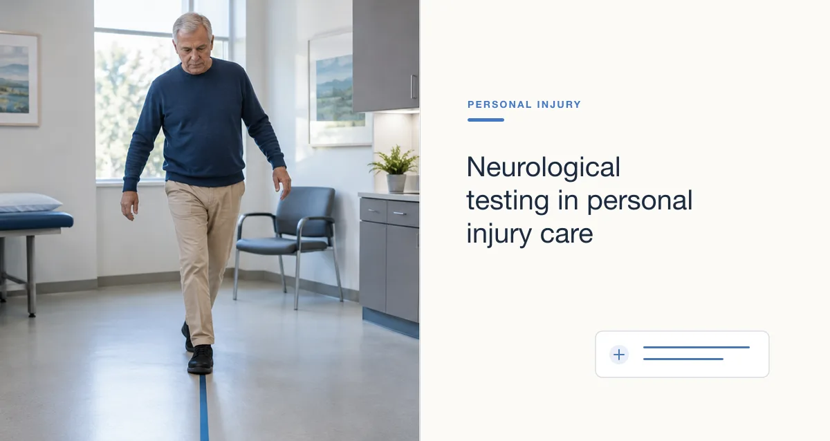
After a car crash or a fall, a patient may describe headaches, numbness, weakness or trouble concentrating. Neurological testing is how a clinician works out what is behind those complaints. In a personal injury case, the same test results are later read by an attorney, an insurance adjuster and sometimes an outside reviewer.
This guide is for neurologists, pain management and spine practices, and the administrators who manage their records. It explains the main tests used after an accident, what each can and cannot show, and how to document results so a non-clinical reader can follow them. Which tests a patient needs is a clinical decision for the treating physician.
Which neurological tests are used after an accident?
The common tools are the neurological examination, electrodiagnostic testing (electromyography and nerve conduction studies), brain and spine imaging, neuropsychological testing, and structured concussion assessment. Clinicians usually combine several, because no single test answers every question.
The National Institutes of Health notes that multiple measures are often used together to diagnose traumatic brain injury (TBI). The same is true for nerve and spine injuries. The table below summarizes what each test is for and where its limits are.
| Test | What it can show | Limits to keep in mind |
|---|---|---|
| Neurological examination | Strength, reflexes, sensation, coordination, cranial nerve function and mental status at a point in time | Findings depend on the examiner and the day; repeating the same elements at each visit makes change visible. |
| Glasgow Coma Scale (GCS) | Level of consciousness from eye, verbal and motor responses; 13 or higher suggests mild TBI | Mostly an early, acute measure; a normal score does not rule out concussion symptoms later. |
| Electromyography (EMG) and nerve conduction studies (NCS) | How muscles and nerves are working; can help separate nerve injury, nerve entrapment and muscle disease | Abnormal results have many possible causes, and results are interpreted with the history and exam. |
| Computed tomography (CT) | Bleeding, bruising or other structural damage, quickly | Often normal in mild TBI; CDC advises against routine imaging to diagnose mild TBI in children. |
| Magnetic resonance imaging (MRI) | More detailed images of the brain and spine, usually in follow-up | Takes longer than CT; findings still have to be correlated with symptoms and the exam. |
| Neuropsychological testing | Memory, attention, processing and other thinking skills measured with standardized tasks | Results can be affected by pain, sleep, mood and effort; interpretation needs a qualified professional. |
| Concussion assessment tools | Symptoms, injury details and risk factors, using a structured form such as CDC's Acute Concussion Evaluation (ACE) | Symptom-based; symptoms can appear hours or days after the injury and change over time. |
Why are concussions hard to document?
Concussion is diagnosed mainly from the clinical picture, not from a scan. Symptoms may not appear right away, they change during recovery, and imaging is often normal. The record has to carry the story through dated symptom reports and repeated exams.
The Centers for Disease Control and Prevention (CDC) groups concussion symptoms into four areas: physical, thinking and remembering, social or emotional, and sleep. CDC also notes that some symptoms show up hours or days later, and that most people feel better within a couple of weeks. When symptoms last longer, the chart is the only place that shows when each one started and how it changed.
- Record the mechanism of injury and the date, in the patient's own words where possible.
- Use one validated symptom scale and repeat it at each visit.
- Note when each symptom first appeared, not only whether it is present.
- Record why imaging was or was not ordered, so a normal or absent scan is not misread.
What do EMG and nerve conduction studies add?
Electrodiagnostic testing shows whether a nerve or muscle is working normally and can help locate a problem, such as a pinched nerve root or an entrapped nerve. It supports a diagnosis built from the history and exam; it does not replace them.
The American Association of Neuromuscular & Electrodiagnostic Medicine (AANEM) states that the physician should review the referral and take a focused history and exam before choosing which nerves and muscles to test. Its position is that nerve conduction studies and needle EMG should usually be done together in the same setting, because either one alone can miss findings. AANEM also says reports should list the nerves tested and the measured values, and that waveforms should be kept in the record when feasible.
For more on charting these results, including structured fields and imaging, see neurology EHR data: diagnostics and documentation.
How should test results be documented for an outside reader?
Each result should state what was tested, why it was ordered, what it showed, and what the treating physician concluded from it. An attorney or adjuster should not have to guess how a test connects to the patient's complaints or to the treatment that followed.
Many personal injury cases take months to resolve, and providers may change along the way. A consistent structure keeps the record readable for anyone who picks it up later:
- Indication: the symptoms and exam findings that led to the test.
- Test details: date, test type, and what was examined.
- Result: the findings, including normal results.
- Interpretation: the physician's impression, in plain language as well as clinical terms.
- Plan: what the result changes, such as a referral, treatment or repeat testing.
Avoid drawing legal conclusions in the note. The physician documents findings and clinical opinion; how a case is valued or decided is not part of the medical record. The same discipline applies to procedures; see how to document injections for an injury case.